ULTRASOUND EXAMINATIONS -
MODERN DIAGNOSTICS AT STEPHANSPLATZ
Ultrasound examination - also known as sonography - is an imaging treatment procedure for visualizing the abdominal organs, thyroid gland, joints and muscles, as well as soft tissues and vessels. The examination is completely painless and risk-free for the patient. An ultrasound examination of the arteries can, for example, detect constrictions and calcifications of the carotid or leg arteries. If detected at an early stage, serious health consequences such as heart attacks or strokes can be avoided in many cases. Ultrasound procedures are also used specifically on the heart to detect constrictions and dilatations of the heart valves and to determine the blood flow there. Thromboses can be detected by this examination method, as can vascular bulges (aneurysms). Doppler and Duplex can also be used to monitor the condition after vascular surgery.
The following examinations are performed in our cardiovascular clinic Esplanade:
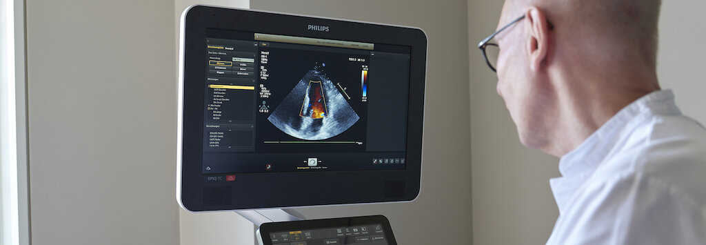
Ultrasound examination of the heart
(echocardiography)
Echocardiography is routine in the diagnosis of heart disease. Doctors also speak of heart ultrasound or heart echo.
Swallowing ultrasound examination of the heart
(Transesophageal Echocardiography/TEE)
TEE stands for transesophageal echocardiography. In this echocardiogram, the examination is not performed from the outside, but through the esophagus.
Stress echocardiography
(Ultrasound examination of the heart under stress).
Stress echocardiography runs like a cardiac echo from the outside, but the heart is additionally challenged. This is done either by medication, or you ride an ergometer during the examination.
Ultrasound examination (color duplex sonography) of the arteries supplying the brain
Color duplex sonography of the arteries supplying the brain is a special ultrasound procedure that can detect narrowing (stenosis) or occlusion of the blood vessels supplying the brain. In addition, we can detect deposits in the vessels.
Ultrasound examination (color duplex sonography) of the aorta, arm, pelvic and leg arteries.
Duplex sonography is a routine method in the modern diagnosis of vascular diseases. In principle, it is performed like any other ultrasound examination. A gel is first applied to the skin of the corresponding body part before the transducer is used.


